The Department of Experimental Physics of Complex Systems at the Institute of Nuclear Physics Polish Academy of Sciences in Kraków, Poland (IFJ PAN) with its own laboratory focuses on research in the low and high dose radiation influence to biological systems. The special interest is in individual radiation sensitivity (its association with human health and cancer occurrence increase), dose response relationships (radiation qualities) and biological dosimetry. The section of Cytogenetic and Molecular Methods uses techniques for dose assessment immediately following exposure to ionizing radiation as different cytogenetic assays: dicentric in a metaphase, micronucleus (MN) in a binucleated cell, premature chromosome condensation (PCC) in an interphase cel, comet assay. The Department has an access to Philips X-ray machine model MCN 323 at 250 kV, 10 mA (own by Department), Hamamatsu X-ray Tube (2 µm beam spot, 160 kV, Ti, Cu, Mo, and W anodes, own by Department), Van de Graaff High Voltage 2.5MV accelerator (own by Department), and 60-230MeV, AIC-144 isochronous cyclotron and Proteus C-235 cyclotron facilities. It is expected that studies and analyses with using X-rays and proton beam will enable the introduction of a relatively simple and rapid predictive test for pre-clinical diagnostics as a rapid test-system for a personal diagnosis of human susceptibility to ionizing radiation. The Cytogenetic and Molecular Methods section has profound experience in interdisciplinary collaborations, needed in molecular methods with expert knowledge in organizing and supervising of the sampling of biological probes and in storing of the biological material. The laboratory is fully equipped in cell culture equipment (cell culture flow cabinets, incubators, centrifuges, light microscopes, semi-automated computerized systems for scoring of chromosome aberrations in: CA, FISH, PCC and MN techniques (MetaSystems, Germany). In the molecular part expression of genes and SNP might be measured with using of RT-PCR machine (ThermoFisher Scientific). In addition the Department is conducting research, using physical methods, for the purpose of eradication cancer and other pathologies. This includes spectroscopic imaging of cells and tissues (examining the internal structure of cells, the cytoskeleton organization, its mechanical and biochemical properties) as well as research at the molecular level.
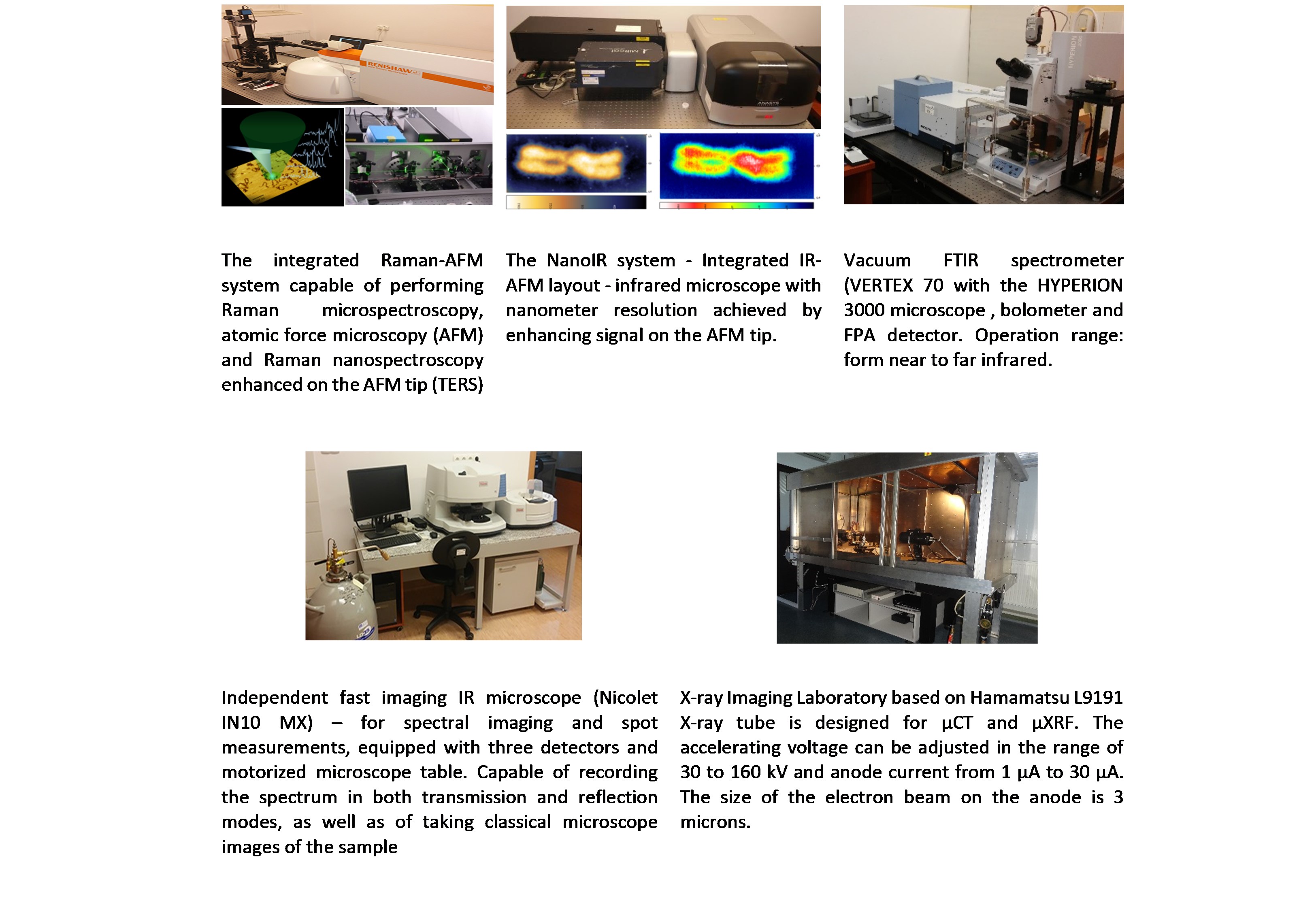
In addition the Department provides opportunity to analyze samples with the use of the proton microprobes with the beam energy of 2 MeV an the intensity of ~1 nA. Rectangular areas (250 x 250 µm2, 128 x 128 pixels) is scanned, with the proton beam of ~20 µm in diameter. Samples are mounted on a precision sample holder (four degrees of freedom) and the beam interaction location is chosen with the use of the long distance microscope system. The corresponding μPIXE and µRBS spectra are registered with the multiparameter, analog data acquisition system working in a single event mode, delivering elemental maps showing distribution of sample elements. The attenuation filters may be used in the X-ray track as required. A parallel, entirely digital data acquisition system has been used to provide high performance X-ray data spectra used for elemental concentration calculations. Examplary equipment and applications are presented below:
Project co-financed by the European Regional Development Fund under the Małopolska Regional Operational Programme for 2007-2013